
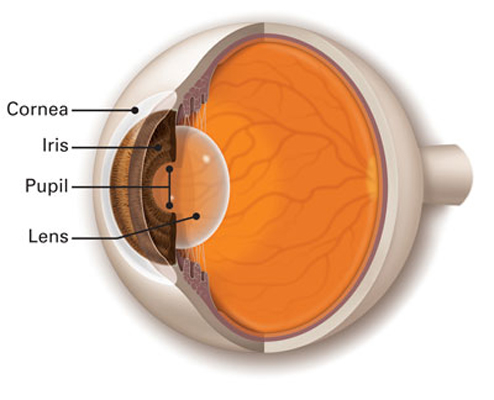
The clear, dome-shaped window of the front of your eye. It focuses light into your eye.
A corneal abrasion is a scratch, scrape on the surface of your cornea. Fingernails, makeup brushes, and tree branches are common culprits of corneal abrasions. Some other causes of corneal abrasion are rubbing your eye and having very dry eyes.
The cornea has many nerve cells. Cells called pain receptors transmit pain to tell us about possible damage to the eye’s surface. In fact, there are hundreds of times more pain receptors in our cornea than there are in our skin.
Your ophthalmologist will put dye called fluorescein on your eye’s surface. Then they will look at your cornea with an instrument called a slit lamp. The dye will highlight a cut or scratch on the cornea.
Your ophthalmologist will treat your eye based on what they find in the exam. The following are some options.
If your corneal abrasion is small, it probably will heal in 1–2 days. A larger corneal abrasion may take about a week to heal.
Corneal dystrophies are a group of rare genetic eye disorders. With corneal dystrophies, abnormal material builds up in the cornea (the clear, front window of the eye). Most corneal dystrophies affect both eyes. They progress slowly and run in families.
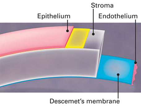
The cornea has five layers:
Corneal dystrophies are caused by the build-up of foreign material in one or more of the five layers of the cornea. The material may cause the cornea to lose its transparency. This can cause loss of vision or blurred vision.
There are more than 20 different types of corneal dystrophies. They are generally grouped into three categories:
The symptoms of Corneal Dystrophy depend upon the type of Corneal Dystrophy. Some people experience no symptoms. In others, the build-up of material in the cornea causes it to become opaque (not clear). This leads toward blurred vision or vision loss.
Many people also experience corneal erosion. This happens when the layer of cells on the surface of the cornea (the epithelium) loosens from the layer underneath (Bowman’s membrane). Corneal erosion causes:
Because most corneal dystrophies are genetic, family history of the disease increases your risk.
Corneal dystrophies can appear at any age. Men and women are equally affected by most corneal dystrophies, except for Fuchs’ dystrophy. Fuchs’ affects women more frequently than men.
Corneal erosion is when the layer of cells on the surface of the cornea, called the epithelium, loosens from the layer underneath. This is painful and makes your vision blurry or hazy.
Corneal erosion pain may start suddenly, often when you first wake in the morning. Your eyes get dry while you sleep, and your eyelid might stick to the cornea. If the epithelium is not firmly attached, opening your eyelids might peel the epithelium off.
The cornea has many nerve cells. Cells called pain receptors transmit the pain to tell us about possible damage to the eye’s surface. In fact, there are hundreds of times more pain receptors in our cornea than there are in our skin.
You are more likely to have corneal erosion if you:
Your ophthalmologist will treat your eye based on what they find in the exam. The following are some options.
The cornea is the clear front window of the eye. A corneal laceration is a cut on the cornea. It is usually caused by something sharp flying into the eye. It can also be caused by something striking the eye with significant force, like a metallic hand tool. A corneal laceration is deeper than a corneal abrasion, cutting partially or fully through the cornea. If the corneal laceration is deep enough it can cause a full-thickness laceration. This is when the laceration cuts completely through the cornea and causes a ruptured globe, a tear into the eyeball itself.
A corneal laceration is a very serious injury and requires immediate medical attention to avoid severe vision loss.
If your eye has been injured, you should do the following:
The cornea is the clear, front window of the eye. It helps focus light into the eye so that you can see. The cornea is made of layers of cells. These layers work together to protect your eye and provide clear vision.
Your cornea must be clear, smooth and healthy for good vision. If it is scarred, swollen, or damaged, light is not focused properly into the eye. As a result, your vision is blurry or you see glare.
If your cornea cannot be healed or repaired, your ophthalmologist may recommend a corneal transplant. This is when the diseased cornea is replaced with a clear, healthy cornea from a human donor.
A human donor is someone who chooses to donate (give) his or her corneas after their death to people who need them. All donated corneas are carefully tested to make sure they are healthy and safe to use.
There are different types of corneal transplants. In some cases, only the front and middle layers of the cornea are replaced. In others, only the inner layer is removed. Sometimes, the entire cornea needs to be replaced.
A corneal ulcer is an open sore on the cornea. The cornea covers the iris and the round pupil, much like a watch crystal covers the face of a watch. A corneal ulcer usually results from an eye infection, but severe dry eye or other eye disorders can cause it.
Symptoms of corneal ulcers include:
See your ophthalmologist immediately if you think you have a corneal ulcer or have any eye symptoms that concern you. Corneal ulcers can badly and permanently damage your vision and even cause blindness if they are not treated.
MBBS, MS FCRS, ICO Consultant